Unexpectedly abnormal PICNIR test and brain SPECT even in the grandson of a patient with dementia
Authors:
A. Bartoš 1; D. Do 2; R. Píchová 2
Authors‘ workplace:
Neurologická klinika 3. LF UK a FN Královské Vinohrady, Praha
; Klinika nukleární medicíny 3. LF UK a FN Královské Vinohrady, Praha
2
Published in:
Cesk Slov Neurol N 2023; 86(6): 405-408
Category:
Letter to Editor
doi:
https://doi.org/10.48095/cccsnn2023405
Dear Editor,
We would like to share unexpected and unique findings in the Picture Oriented Picture Naming and Equipment (PICNAV) test and single-photon emission computed tomography (SPECT) of the brain even in the grandson of a former patient with dementia, which are unprecedented in the world [1-4]. Cognitive changes on POBAV and Amnesia Light and Brief Assessment (ALBA) tests and hypoperfused precincts on brain SPECT were observed not only in a young patient with a genetically proven cause of dementia, but even in the daughter of one of our patients with dementia [5,6]. It is not yet known whether any changes also occur in persons two generations younger, i.e. grandchildren of patients with dementia. Surprisingly, because we caught them at such a young age, we present the abnormal findings in the following case report, which takes these readily available methods to new heights.
Between 2010 and 2016, a grandson repeatedly accompanied his grandmother from the age of 74 to our neurological outpatient clinic for memory disorders at the Neurological Clinic of the University Hospital Královské Vinohrady (FNKV) and the 3rd Medical Faculty of Charles University in Prague. The combined disease Alzheimer's disease with frontotemporal dementia progressed from mild cognitive impairment to dementia and finally to death.
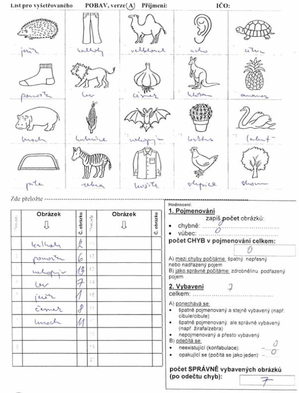
Five years after the end of the patient's follow-up in the outpatient clinic and one year after her death, the 30-year-old grandson requested a preventive cognitive examination because he observed an increasing difficulty in remembering and recalling information. The college-educated individual wondered if this was a completely natural process or even the fault of modern living and the overuse of technology. In the third version of the Addenbrooke's Cognitive Examination (ACE-III), he scored a normal score with almost a full 97 out of 100. He lost only 3 points in word production to the letter P, listing only 10 words in one minute. At the same time, he was examined by our own cognitive tests. In one of our battery of tests he lost only 2 points out of a maximum of 35 points. This included a gesture test (TEGEST) with a score of 6 correctly presented gestures, all of which were equipped with immediate and remote [1,2]. In our other home battery of short tests, he scored 30 out of a maximum of 35 points. This included a hedgehog version of the POBAV test, in which he scored as follows: 0 naming errors // 7 correctly equipped pictures (Figure 1). Although the POBAV test norms for this age group are unknown, given his young age and high education, we would have expected better performance in the second test battery and especially in the POBAV test. According to Czech performance on the hedgehog version of the POBAV test, more educated older persons (with a high school diploma or more or 15 years of education or more) should be able to recall at least 7 correct picture names [1]. This number of picture names is borderline for older individuals. It is exactly the result achieved by an individual two generations younger. The recording form and instructions for using the POBAV test are freely downloadable from the ABADECO website [7].


Because of subjective complaints of memory impairment, poorer P-word production and the findings in the POBAV test, he accepted the offer of a functional brain examination at the Nuclear Medicine Clinic at the National University of Health Sciences.
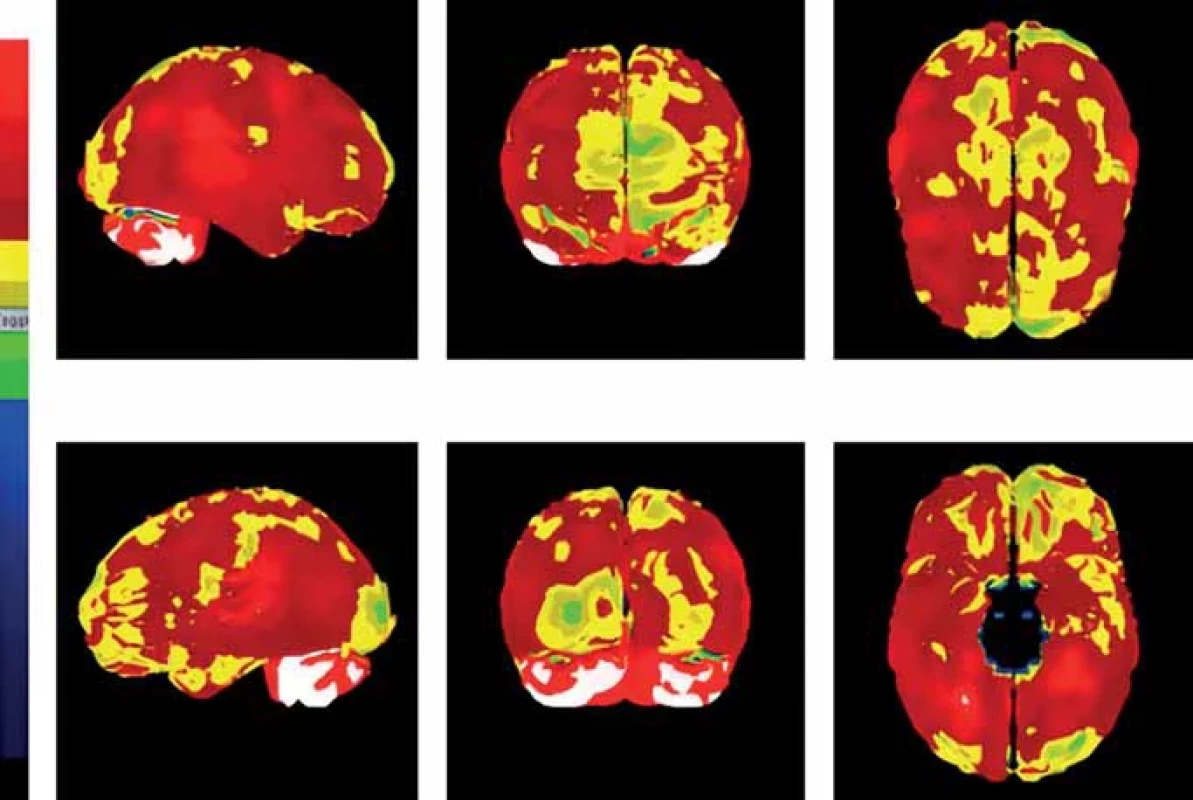
The examination of regional brain perfusion by SPECT technique was performed according to the protocol of the European Association of Nuclear Medicine [8]. The subject was i.v. injected with the radiopharmaceutical 99mTc-HMPAO (hexa-methyl-propylene-amino-oxime) while lying supine in a quiet room with eyes closed. After 30 min, brain perfusion images were recorded using SPECT technique on an Infinia-HAWKEYE gamma camera (GE HealthCare, Chicago, IL, USA) with a fan-beam collimator, image reconstruction was performed using an iterative technique, and images of sections in three planes (coronal, transverse and sagittal) including three-dimensional images were obtained. The reconstructed data were then processed using NeuroGam software for semi-quantitative assessment of cerebral perfusion.
In our patient, hypoperfusion was not evident on conventional transverse, coronal and sagittal sections. After quantification with projection of hypoperfused regions onto a three-dimensional brain model, regions with lower perfusion were more evident (Figure 2). Activity accumulation was slightly inhomogeneous. Borderline to mild reductions in perfusion were most diffuse in the left hemisphere, but mainly frontal, parietal, and occipital. The perfusion of the examined person was compared to the normal Segami NeuroGam database corresponding to the age category 16-45 years, not taking into account gender.
This finding was so surprising and disturbing to the nuclear medicine doctors that they first looked for a flaw in the actual performance of the examination. A review of the procedures found no fault with the staff or the technical execution. The possibility of unwanted head movement was considered, and thus software correction for movement was applied in the reconstruction of the images. After several days of doubt, deliberation and numerous re-constructions of the data, the resulting images were the same. Thus, a re-reading by the physician including evaluation with time intervals of several hours and days was also proceeded. And the result was still the same.
The result of the examination was then gently consulted with the grandson and a healthy lifestyle was recommended. He himself summed it up in words that illustrate his own embarrassed feelings: 'Thank you for the report. The low activity left doesn't comfort me much, so I'll definitely check back in a few years. In the meantime, I also keep my fingers crossed that you do well, both in your research and in your personal life." We consider it important to continue to monitor our grandson at least using the POBAV and SPECT methodologies used. Follow-up examinations may give an answer as to whether these were non-specific findings and fluctuations or will be consistent in nature over time. Otherwise, he had normal brain MRI and blood findings except hyperbilirubinemia known since childhood.
This first world description and findings open questions and further perspectives. Does the individual already have established brain changes at such a young age that predispose him to a similar fate as his grandmother decades later? An initial search for individuals at risk of developing cognitive impairment could be performed using the very brief yet challenging ALBA and POBAV tests in person or remotely online [5,6,9]. In suitable candidates, early changes could be demonstrated by brain SPECT scans, enhanced by quantification and subsequent three-dimensional visualization of cerebral perfusion. This is performed using a semi-quantitative program such as NeuroGam, which greatly enhances the information potential of functional imaging compared to single plane images, as demonstrated in our case report. Semiquantitative assessment could become a tool to recognize perfusion dysfunction in the earlier stages of dementia, when the typical pattern is not yet sufficiently expressed visually. Although hypoperfusion may be nonspecific, its finding increases diagnostic accuracy.
A short video on YouTube explains for the interested reader how to properly process tomographic brain sections into three-dimensional brains using Neurogam [10]. With its more frequent use, we can see interesting findings not only in patients with incipient cognitive deficits, but also in the offspring of patients with dementia.
Grant support
This work was supported by the Charles University Neuroscience COOPERATIO project and the Czech Ministry of Health - RVO (FNKV, 00064173).
Conflict of interest
The authors declare that they have no conflict of interest in relation to the subject of the paper.
This is an unauthorised machine translation into English made using the DeepL Translate Pro translator. The editors do not guarantee that the content of the article corresponds fully to the original language version.
Sources
1. Bartoš A. Praktický návod k identifikaci zapomětlivého pacienta podle kognitivních testů Amnesia Light and Brief Assessment (ALBA) a Pojmenování obrázků a jejich vybavení (POBAV) k velmi rychlému vyšetření nejen paměti. Geri a Gero 2022; 11 (3): 118–128.
2. Bartoš A. Inovativní a původní české kognitivní testy Amnesia Light and Brief Assessment a Pojmenování obrázků a jejich vybavení. Medicína pro praxi 2022; 19 (1): 50–57. doi: 10.36290/med.2022.007.
3. Pichova R, Bartoš A, Lang O. SPECT mozku u kognitivních poruch – porovnání s klinickou diagnózou a význam pro klinickou praxi – stále platná možnost? Nukl Med 2022 : 11 : 42–48.
4. Ferrando R, Damian A. Brain SPECT as a biomarker of neurodegeneration in dementia in the era of molecular imaging: Still a valid option? Front Neurol 2021; 12 : 629442. doi: 10.3389/fneur.2021.629442.
5. Nosková E, Bartoš A, Kopeček M. Od diagnózy deprese, generalizované – úzkostné poruchy, schizoafektivní poruchy až k diagnostice presenilní demence spojené s mutací proteinu tau asociovaném s mikrotubuly (MAPT) S305N aneb slabiny fenomenologické klasifikace. Psychiatr praxi 2022; 23 (1): 20–29. doi: 10.36290/psy.2022. 004.
6. Bartoš A. ALBA and PICNIR tests used for simultaneous examination of two patients with dementia and their adult children. Ces Slov Neurol N 2021; 84/117 (6): 583–586. doi: 10.48095/cccsnn2021583.
7. Vizuální škály HIP-HOP a PAS na CT/MR mozku. [online]. Dostupné z URL: http: //www.abadeco.cz.
8. Kapucu OL, Nobili F, Varrone A et al. EANM procedure guideline for brain perfusion SPECT using 99mTc-labelled radiopharmaceuticals, version 2. Eur J Nucl Med Mol Imaging 2009; 36 (12): 2093–2102. doi: 10.1007/s00259-009-1266-y.
9. Polanská H, Bartoš A. Telemedicínské vyšetření kognitivní testy ALBA, POBAV a ACE-III. Ces Slov Neurol N 2022; 85/118 (4): 296–305. doi: 10.48095/cccsnn2022296.
10. Zpracování tomografických řezů mozku do třírozměrných mozků pomocí programu Neurogam. [online]. Dostupné z URL: https://www.youtube.com/ watch?v=Ke8oQEyEWB4.
Labels
Paediatric neurology Neurosurgery NeurologyArticle was published in
Czech and Slovak Neurology and Neurosurgery

2023 Issue 6
-
All articles in this issue
- Surgical treatment of intracranial aneurysm recurrence after clipping
- Diffuse glioma overview based on the 2021 WHO classification part 1 – adult type
- The effect of chemotherapy on cognitive functions in children with leukemia
- Treatment of sleep disorders with repetitive transcranial magnetic stimulation
- The effects of introducing psychoeducational programs in patients with stroke in post-acute care
- Memory reserve and memory maintenance in SuperAgers
- Polysomnographic findings in men over 55 years of age with narcolepsy type 1
- Z. Adam et al. Monoklonální gamapatie klinického významu a další nemoci
- Zpráva o výročním sjezdu České neurochirurgické společnosti ČLS JEP v Hradci Králové
- Atypical cases of cycloplegia caused by Datura stramonium
- Unexpectedly abnormal PICNIR test and brain SPECT even in the grandson of a patient with dementia
- Czech and Slovak Neurology and Neurosurgery
- Journal archive
- Current issue
- About the journal
Most read in this issue
- Diffuse glioma overview based on the 2021 WHO classification part 1 – adult type
- Treatment of sleep disorders with repetitive transcranial magnetic stimulation
- The effects of introducing psychoeducational programs in patients with stroke in post-acute care
- Surgical treatment of intracranial aneurysm recurrence after clipping
