
Intraoperačná spinálna sonografia – potenciálny prínos pri degeneratívnych ochoreniach chrbtice
Published in:
Cesk Slov Neurol N 2026; 89(5): 327-333
Category:
Short Communication
doi:
https://doi.org/10.48095/cccsnn2026aop01
Overview
While the modernization of ultrasound devices and the development of miniature probes since the turn of the millennium have improved the visualization of both normal and pathological structures within the spinal canal, the number of publications dedicated to intraoperative spinal sonography in the context of degenerative spinal disorders remains low. Although rarely utilized, intraoperative ultrasound (IOUS) can provide valuable information during surgery for various degenerative spinal pathologies in some cases. In this short communication, we present four illustrative cases that highlight the potential benefits of intraoperative spinal sonography: one case demonstrating the use of IOUS during a decompressive cervical laminectomy, one case illustrating the benefit of IOUS during the excision of a thoracic disc herniation compressing the spinal cord, and two cases of lumbar microdiscectomy performed using IOUS. In each case, sonographic visualization facilitated the course of the surgical procedure to some extent. While spinal IOUS is not an indispensable tool in these diagnoses, in selected cases of degenerative spinal disorders it can help prevent unnecessary dissection, or, conversely, demonstrate that the decompression of neural structures remains suboptimal. Further research is needed to establish the definitive role of IOUS in the surgical management of degenerative spinal disorders.
Keywords:
Spinal cord – magnetic resonance imaging – disc herniation – intraoperative ultrasound – spinal roots
Introduction
While intraoperative ultrasound (IOUS) has become a relatively established tool in cranial surgery over the past decade, its application in spinal procedures remains more limited, and is predominantly reserved for tumorous lesions [1,2]. The use of IOUS in the context of degenerative spinal disorders is far less common; however, it can serve as a valuable adjunct in selected cases [3]. The aim of this short communication is to present four illustrative cases in which the utilization of IOUS positively influenced the surgical course.
Illustrative case 1
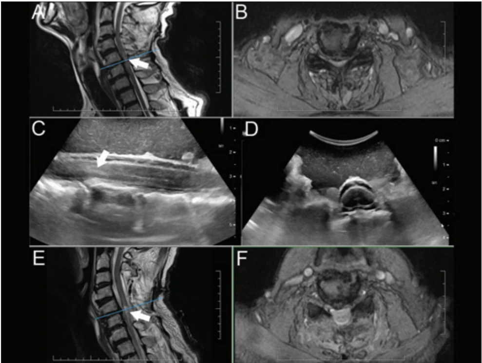
A 73-year-old male with a complex medical history including colon cancer with hepatic metastases, liver cirrhosis, esophageal varices, ischemic heart disease and arterial hypertension conveying high cardiovascular risk suffered a fall and subsequently developed paraplegia and severe upper limb paresis, accompanied by sensory loss extending distally from the nipple line. MRI revealed primary cervical spinal canal stenosis, multi-level disc bulging, and compressive myelopathy at the C5–C6 level (Fig. 1). Intravenous corticosteroid therapy and an urgent decompressive laminectomy were indicated. Following a C3–C6 laminectomy, an IOUS scan (BK5000 ultrasound system with a BK N13C5 craniotomy transducer, BK Medical Aps, Herlev, Denmark) demonstrated an adequate extent of spinal cord decompression (Fig. 1), thereby obviating the need for further manipulation near the spinal cord. However, the hyperechogenic focus within the spinal cord appeared slightly more prominent than expected based on the preoperative MRI, predicting a complicated neurological recovery (Fig. 1). Postoperative MRI confirmed the ultrasound findings. In the postoperative period, the patient demonstrated only mild neurological improvement: sensation was preserved across his upper extremities, trunk, and lower extremities, and motor function in his upper extremities improved bilaterally in all segments with the exception of finger flexion and extension. The planned posterior stabilization was postponed due to an acute, severe COVID-19 infection.
Illustrative case 2
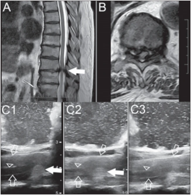
A 74-year-old male with a history of arterial hypertension and chronic lymphocytic leukemia presented with a five-week history of lower back pain radiating to the lower limbs and progressive paraparesis. On admission, he was non-ambulatory with absent tactile sensation in the lower extremities. MRI of the thoracic spine revealed a right-sided lateral disc herniation and spinal stenosis at the T10-11 level, causing severe spinal cord compression. A posterior decompressive T10 laminectomy was indicated. The herniated thoracic disc was approached lateral to the dura (without the need for dural manipulation) via a posterior transfacet pedicle-sparing approach, as described by Nishimura and colleagues [4]. Following a seemingly adequate discectomy, IOUS (using a BK X18L5s hockey stick transducer) revealed persistent spinal cord compression caused by a residual disc herniation; the remaining disc material was subsequently removed piecemeal (Fig. 2). Postoperatively, the patient was successfully mobilized, regaining ambulatory status, and reported improvement of the sensation in his lower extremities.
Illustrative case 3
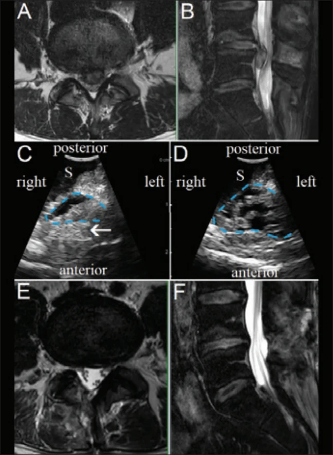
A 47-year-old male with a history of arterial hypertension presented with severe bilateral radicular pain radiating down the posterior aspects of the lower limbs, accompanied by severe bilateral peroneal nerve paresis of three weeks‘ duration. The patient had previously undergone repeated spinal ozone therapy at an outside facility, without clear clinical benefit. Preoperative MRI revealed a massive, caudally migrated medial disc extrusion at the L4–L5 level. An urgent decompression and discectomy were indicated. Intraoperatively, the surgical approach was complicated by dense epidural adhesions. Given the large medial nature of the herniation and the bilateral symptomatology, there was a preoperative discussion regarding the optimal side for the hemilaminectomy to achieve sufficient contralateral decompression. Following the microdiscectomy via a right-sided partial L4 and L5 hemilaminectomy, IOUS was utilized to evaluate the spinal canal using a miniature ultrasound probe (BK N11C5s “burr hole” transducer). The IOUS clearly demonstrated adequate decompression of the contralateral side, obviating the need for further surgical exploration using curved microdissectors (Fig. 3). Postoperatively, the patient was successfully mobilized, reporting significant relief of his bilateral radicular pain, and the paresis fully resolved.
Illustrative case 4
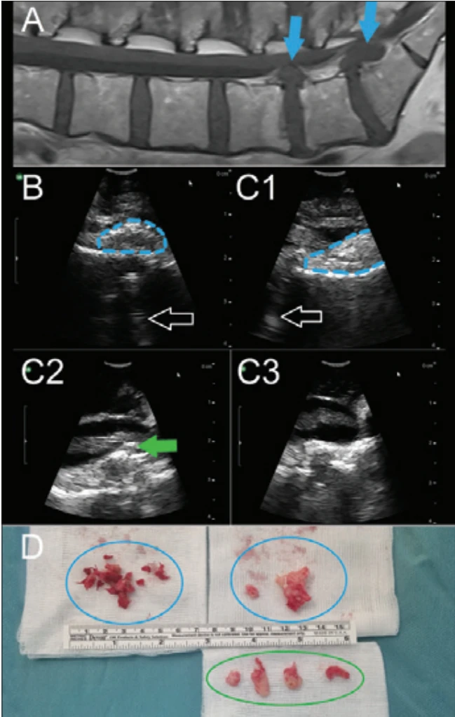
A 50-year-old female, who had initially received care abroad following multiple prior administrations of ozone therapy, was admitted with severe left-sided S1 radiculopathy and cauda equina syndrome of four days‘ duration. This manifested as saddle anesthesia and urinary retention (requiring urinary catheter placement at the referring facility). Preoperative MRI revealed a voluminous disc extrusion at L5–S1 and a smaller herniation at L4–L5. She underwent an urgent left-sided hemilaminectomy at L5 with a partial hemilaminectomy at L4 and S1. After the L4–L5 discectomy, dense epidural adhesions were encountered at the L5–S1 level – the S1 root was unidentifiable, and the dural sac was severely adherent to the ventral spinal canal. Following the initial extraction of a large sequester, IOUS using a miniature probe (BK N11C5s “burr hole” transducer) revealed a relatively large residual herniation fragment that had been previously obscured by the adhesions (Fig. 4). This fragment was subsequently identified and removed in four smaller pieces. In the immediate postoperative period, the patient experienced significant relief of her radicular pain, though her sphincter deficits initially persisted. At the one-year postoperative follow-up, the patient was fully ambulatory, reporting mild residual numbness in the left S1 dermatome, intact bowel control, and the ability to sense bladder filling and urinate voluntarily, although occasional stress incontinence occurred during heavy physical exertion.
Discussion
The first report addressing the intraoperative use of ultrasound in spinal surgery was published by Reid in 1978 [5], describing the ultrasound visualization of a cervical spinal cord tumor. In the 1980s, in addition to ultrasound-guided procedures for neoplastic spinal lesions, utilization of IOUS during surgery for degenerative disorders such as lumbar disc herniation was described [6,7]. Shortly thereafter, intraoperative ultrasound-guided surgeries for both neoplastic and degenerative spinal disorders were described in the former Czechoslovakia by a group led by Prof. Fagula and Prof. Šulla [8,9]. Throughout the 1980s and 1990s, the quality of ultrasound imaging was insufficient for adequate visualization of the relatively delicate structures of the spinal canal.
After the turn of the millennium, improvements in the quality of ultrasound systems and the miniaturization of ultrasound probes led to more frequent use of intraoperative spinal ultrasonography during tumor surgeries [1,10,11]. Although IOUS is currently utilized far more frequently for neoplastic lesions of the spine and spinal cord than for degenerative pathologies, it can serve as a helpful adjunct in selected cases of degenerative disorders as well [3].
First, it may be used to verify the extent of spinal cord decompression in patients with cervical spinal stenosis and myelopathy [12–15]. In these cases, ultrasound not only displays the extent of decompression (and thus its sufficiency) but may also provide intraoperative prognostic indications regarding postoperative neurological recovery. Chen and colleagues [16] found that the extent of spinal cord hyperechogenicity after decompression significantly correlated with the postoperative functional status of the spinal cord (i.e., the likelihood of a postoperative neurological deficit). In our case, the appearance and extent of cervical spinal cord hyperechogenicity indicated limited potential for neurological improvement. However, further studies are necessary to assess whether the routine use of IOUS has the potential to improve patient prognosis.
Second, IOUS may be successfully utilized during the resection of thoracic disc herniations, especially via posterolateral approaches [3,4,17–19], where a well-known limitation is the poor visualization of close--to-midline herniations [20]. Advances in IOUS, however, have improved the visualization of ventral pathologies, obviating the need for any significant thecal sac retraction [19]. Recently, the advantages of IOUS utilization during a lateral extracavitary approach in thoracic disc herniation surgery were also described; IOUS was used to confirm complete decompression over the spinal cord and to rule out the presence of unnoticed fragments [20]. In our case 2, who was operated on via a transfacet pedicle-sparing approach, IOUS significantly contributed to a successful surgical course by demonstrating persistent spinal cord compression at a point when an experienced spinal surgeon (J.M.) considered the anterior decompression to be sufficient (Fig. 2). Although traditional thoracic discectomy will likely be replaced by full endoscopic thoracic discectomy in a large proportion of patients – due to the minimally invasive nature of the endoscopic approach – spinal endoscopy is still not available in all centers, and its learning curve is relatively steep [21]. Interestingly, in a recent retrospective multi-center case series and meta-analysis comparing endoscopic approaches with traditional thoracic microdiscectomy, only 41 patients operated on using the endoscopic approach were included (vs. 3,190 patients operated on using traditional thoracic microdiscectomy) [22].
Third, IOUS can offer potential benefits during lumbar microdiscectomies. Although lumbar discectomies are generally considered “simpler” neurosurgical procedures, they still carry a risk of several complications, including inadvertently leaving a resectable sequestered disc fragment in some cases [23–26]. Of note, current IOUS systems appear to possess sufficient imaging quality to enable the visualization of residual lumbar disc material inadvertently left behind, at least in a subset of cases [3]. Aoyama and colleagues [27] examined a cohort of 30 patients with lumbar disc herniation. In two cases, they identified a residual herniated disc that they would not have detected without using ultrasound. Interestingly, dedicated ultrasound miniprobes allow for the visualization of anatomical structures within the spinal canal even during minimally invasive procedures, such as tubular microdiscectomies [28]. New lateral-firing miniprobes can provide striking image quality after insertion into the spinal canal through a small bony window; this approach represents a conceptual shift from conventional extraspinal ultrasound (where a larger bony window is sometimes required) toward an intraspinal imaging paradigm [29]. IOUS may also help establish the intradural location of a lumbar disc herniation, guide the dural opening in these cases and confirm adequate nerve root decompression following discectomy [30]. In both of our illustrative cases of lumbar disc herniation, IOUS provided sufficient imaging of the anatomical structures within the spinal canal (Fig. 3 and 4). In the first lumbar case (case 3), IOUS showed sufficient decompression of the cauda equina nerve roots with no detectable residual disc fragment. In the second patient (case 4), who presented with cauda equina syndrome caused by a two-level lumbar disc herniation and significant epidural fibrosis (likely due to repeated ozone therapy [31]), IOUS helped identify a relatively large residual herniation fragment at the L5–S1 level (compressing the S1 nerve root) that had been previously obscured by the adhesions. The fragment was subsequently located and extracted successfully. Of note, while IOUS is certainly not an indispensable tool during lumbar discectomies, intraoperative video documentation of the absence of residual fragments may assist in differentiating true recurrence from incomplete decompression in cases of suspected ultra-early reherniation.
Lastly, while standard two-dimensional IOUS is currently the only surgically relevant modality, it is not the only sonography-based imaging method potentially applicable during spinal surgery. Initial attempts at three-dimensional sonographic imaging of intradural spinal tumors have been published; however, to date, this remains largely an experimental methodology [32], and is not yet a routine modality in spinal surgery. Another potential application of IOUS involves utilizing intraoperative US-based navigation for pedicle screw insertion, relying on automatic registration between IOUS scans and preoperative CT images [33,34]. In the future, this novel method could theoretically serve as a low-cost, radiation-free alternative to intraoperative CT and fluoroscopy.
Conclusions
Although spinal IOUS is not an indispensable tool in surgery for degenerative spinal disorders, in selected cases, it can help prevent unnecessary dissection; or, conversely, demonstrate that the decompression of neural structures remains suboptimal. Further research is needed to establish the definitive role of IOUS in the surgical management of degenerative spinal disorders.
Ethical principles
The study was conducted in accordance with the Declaration of Helsinki (2013 revision). The study protocol was approved by the institutional ethics committee (Etická komisia UN Bratislava, Nemocnica akad. L. Dérera) on May 28, 2025 (approval number: 11/2025). All patients signed an informed consent form for participation in the study and for the publication of their data and images.
Funding
The Scientific Grant Agency of the Ministry of Education of the Slovak Republic and the Slovak Academy of Sciences (VEGA) provided financial support in the form of a grant (number 1/0760/24). The sponsor had no role in the design or conduct of this research.
Conflict of interest
The authors declare they have no potential conflicts of interest concerning drugs, products, or services used in the study.
Sources
1. Moják P, Filip M, Linzer P et al. Intradurálne extramedulárne nádory chrbtice. Cesk Slov Neurol N 2019; 82/115 (2): 125–139. doi: 10.14735/amcsnn2019125.
2. Vetrano IG, Prada F. Intraoperative Findings in Spinal Lesions. In: Prada F, Solbiati L, Martegani A et al (eds). Intraoperative Ultrasound (IOUS) in Neurosurgery. Cham: Springer 2016 : 59–69. doi: 10.1007/978-3-319-25268-1_6.
3. Vasudeva VS, Abd-El-Barr M, Pompeu YA et al. Use ofintraoperative ultrasound during spinal surgery. GlobalSpine J 2017; 7 (7): 648–656. doi: 10.1177/2192568217700100.
4. Nishimura Y, Thani NB, Tochigi S et al. Thoracic discectomy by posterior pedicle-sparing, transfacet approach with real-time intraoperative ultrasonography. J Neurosurg Spine 2014; 21 (4): 568–576. doi: 10.3171/2014.6.SPINE13682.
5. Reid MH. Ultrasonic visualization of a cervical cord cystic astrocytoma. AJR Am J Roentgenol 1978; 131 (5): 907–908. doi: 10.2214/ajr.131.5.907.
6. Knake JE, Gabrielsen TO, Chandler WF et al. Real-time sonography during spinal surgery. Radiology 1984; 151 (2): 461–465.
7. Koivukangas J, Tervonen O. Intraoperative ultrasound imaging in lumbar disc herniation surgery. Acta Neurochir (Wien) 1989; 98 : 47–54
8. Sulla I, Santa M, Fagul‘a J. Two-dimensional ultrasonography during spinal surgery. Zentralbl Neurochir 1990; 51 (3): 160–163
9. Santa M, Sulla I, Fagul‘a J. Peroperative ultrasound examination of the spinal canal. Cesk Slov Neurol N 1991; 54 (1): 6–13.
10. Zhou H, Miller D, Schulte DM et al. Intraoperative ultrasound assistance in treatment of intradural spinal tumours. Clin Neurol Neurosurg 2011; 113 (7): 531–537. doi: 10.1016/j.clineuro.2011.03.006.
11. Prada F, Vetrano IG, Filippini A et al. Intraoperative ultrasound in spinal tumor surgery. J Ultrasound 2014; 17 (3): 195–202. doi: 10.1007/s40477-014-0102-9.
12. Schär RT, Wilson JR, Ginsberg HJ. Intraoperative ultrasound-guided posterior cervical laminectomy for degenerative cervical myelopathy. World Neurosurg 2019; 121 : 62–70. doi: 10.1016/j.wneu.2018.09.217.
13. Tat J, Tat J, Yoon S et al. Intraoperative ultrasound in spine decompression surgery: a systematic review. Spine (Phila Pa 1976) 2022; 47 (2): E73–E85. doi: 10.1097/BRS.0000000000004111.
14. Patel MR, Jacob KC, Parsons AW et al. Systematic review: applications of intraoperative ultrasonography in spinal surgery. World Neurosurg 2022; 164: E45–E58. doi: 10.1016/j.wneu.2022.02.130.
15. Rustagi T, Das K, Chhabra HS. Revisiting the role of intraoperative ultrasound in spine surgery for extradural pathologies: review and clinical usage. World Neurosurg 2022; 164 : 118–127. doi: 10.1016/j.wneu.2022.04.116.
16. Chen G, Wu H, Chen N et al. Potential of intraoperative ultrasonographic assessment of the spinal cord central echo complex in predicting postoperative neurological recovery of degenerative cervical myelopathy. Eur J Neurol 2022; 29 (1): 217–224. doi: 10.1111/ene.15109.
17. Tan LA, Lopes DK, Fontes RBV. Ultrasound-guided posterolateral approach for midline calcified thoracic disc herniation. J Korean Neurosurg Soc 2014; 55 (6): 383–386. doi: 10.3340/jkns.2014.55.6.383
18. Wessell A, Mushlin H, Fleming C et al. Thoracic discectomy through a unilateral transpedicular or costotransversectomy approach with intraoperative ultrasound guidance. Oper Neurosurg (Hagerstown) 2019; 17 (3): 332–337. doi: 10.1093/ons/opy348.
19. Saway BF, Alshareef M, Lajthia O et al. Ultrasonic spine surgery for every thoracic disc herniation: a 43-patient case series and technical note demonstrating safety and efficacy using a partial transpedicular thoracic discectomy with ultrasonic aspiration and ultrasound guidance. J Neurosurg Spine 2022; 36 (5): 800–808. doi: 10.3171/2021.8.SPINE21819.
20. García Pérez F, Gomar Alba M, Guil Ibáñez JJ et al. The role of intraoperative ultrasound during lateral extracavitary approach in thoracic disc herniation surgery. Tips and tricks for lateral approaches. J Ultrasound 2026; 29 : 205–211. doi: 10.1007/s40477-025-01093-w.
21. Park SC, Park SM, Chung HJ et al. Learning curve for full-endoscopic lumbar decompression via interlaminar approach using the learning curve cumulative summation analysis. J Orthop Surg Res 2025; 20 (1): 297. doi: 10.1186/s13018-025-05699-y.
22. Sofoluke N, Leyendecker J, Barber S et al. Endoscopic versus traditional thoracic discectomy: a multicenter retrospective case series and meta-analysis. Neurosurgery 2025; 96 (1): 152–171. doi: 10.1227/neu.0000000000003034.
23. Abdelhamid MS, Elfiky TA, El Saghir HA et al. Significance of intraoperative induced cough reflex in reducing the incidence of residual and recurrent lumbar disc herniation after microdiscectomy. A prospective cohort study. Alex J Med 2025; 61 (1): 171–176. doi: 10.1080/20905068.2025.2456694.
24. Hadgaonkar S, Tomer D. Analogy of lumbar disc: retained, residual, or recurrent disc? J Orthop Case Rep 2023; 13 (12): 01–04. doi: 10.13107/jocr.2023.v13.i12.4052.
25. Baek J, Yang SH, Kim CH et al. Postoperative longitudinal outcomes in patients with residual disc fragments after percutaneous endoscopic lumbar discectomy. Pain Physician 2018; 21 (4): E457–E466.
26. Noh SH, Ndraha E, Shin DA et al. Immediate postoperative lumbar spine magnetic resonance imaging: correlation with postoperative pain in lumbar microdiscectomy. Medicine (Baltimore) 2022; 101 (43): e31287. doi: 10.1097/MD.0000000000031287.
27. Aoyama T, Hida K, Akino M et al. Detection of residual disc hernia material and confirmation of nerve root decompression at lumbar disc herniation surgery by intraoperative ultrasound. Ultrasound Med Biol 2009; 35 (6): 920–927. doi: 10.1016/j.ultrasmedbio.2008.12.014.
28. Tawil ME, Chryssikos T, Dada A et al. Use of minimally invasive ultrasound transducer during tubular microdiscectomy. Neurosurg Focus Video 2024; 10 (2): V6. doi: 10.3171/2024.1.FOCVID23206.
29. Stroop R, Zawy Alsofy S, Nakamura M et al. Feasibility of Intraoperative intraspinal endosonography using a miniaturized ultrasound probe through an extended interlaminar lumbar approach: a pilot study. J Clin Med 2026; 15 (11): 4090. doi: 10.3390/jcm15114090.
30. Wei MC, Castle-Kirszbaum M, Kam JKT. Intraoperative ultrasound and findings of lumbar intradural disk herniation causing cauda equina syndrome. World Neurosurg 2023; 179 : 5–7. doi: 10.1016/j.wneu.2023.07.128.
31. Vanni D, Galzio R, Kazakova A et al. Intraforaminal ozone therapy and particular side effects: preliminary results and early warning. Acta Neurochir (Wien) 2016; 158 (5): 991–993. doi: 10.1007/s00701-016-2755-y.
32. Saß B, Bopp M, Nimsky C et al. Navigated 3-dimensional intraoperative ultrasound for spine surgery. World Neurosurg 2019; 131: e155–e169. doi: 10.1016/j.wneu.2019.07.188.
33. Brignol A, Gueziri HE, Cheriet F et al. Automatic extraction of vertebral landmarks from ultrasound images: a pilot study. Comput Biol Med 2020; 122 : 103838. doi: 10.1016/j.compbiomed.2020.103838.
34. Gueziri HE, Georgiopoulos M, Santaguida C et al. Ultrasound-based navigated pedicle screw insertion without intraoperative radiation: feasibility study on porcine cadavers. Spine J 2022; 22 (8): 1408–1417. doi: 10.1016/j.spinee.2022.04.014.
Labels
Paediatric neurology Neurosurgery NeurologyArticle was published in
Czech and Slovak Neurology and Neurosurgery
2026 Issue 5
Most read in this issue
- Safety aspects of transitioning from alteplase to tenecteplase in the treatment of acute ischemic stroke – a narrative review
- 100 years of the Czech Department of Neurology, Charles UniversityThe fates of Czech and German neurologists in Prague from 1938 to 1945
- Anatomy, etiology, and graphic imaging findings of hypertrophic olivary degeneration – description of 10 cases
- Patterns of cognitive-motor interference in patients with post-stroke cognitive impairment