
Isolated orbital mucocele as a late sequela following skull base fracture – case report and review of the literature
Authors:
J. Šipoš; M. Liška; J. Kozák
Authors‘ workplace:
Neurosurgical department, Slovak Medical University, University Hospital Bratislava
Published in:
Cesk Slov Neurol N 2026; 89(5): 334-337
Category:
Letter to Editor
doi:
https://doi.org/10.48095/cccsnn2026334
Dear Editor,
Mucocele is benign, slow-growing, mucus-filled cavity in the paranasal sinuses caused by inflammation, trauma, surgery or anatomical abnormalities [1]. We report a rare case of isolated orbital mucocele that developed as a late sequela following a skull base fracture. In addition, we reviewed English-language literature using PubMed database to identify relevant articles published through August 2025.
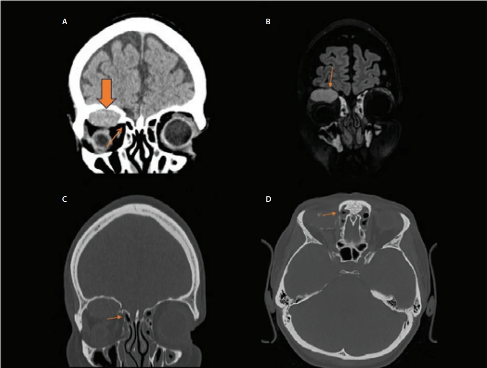
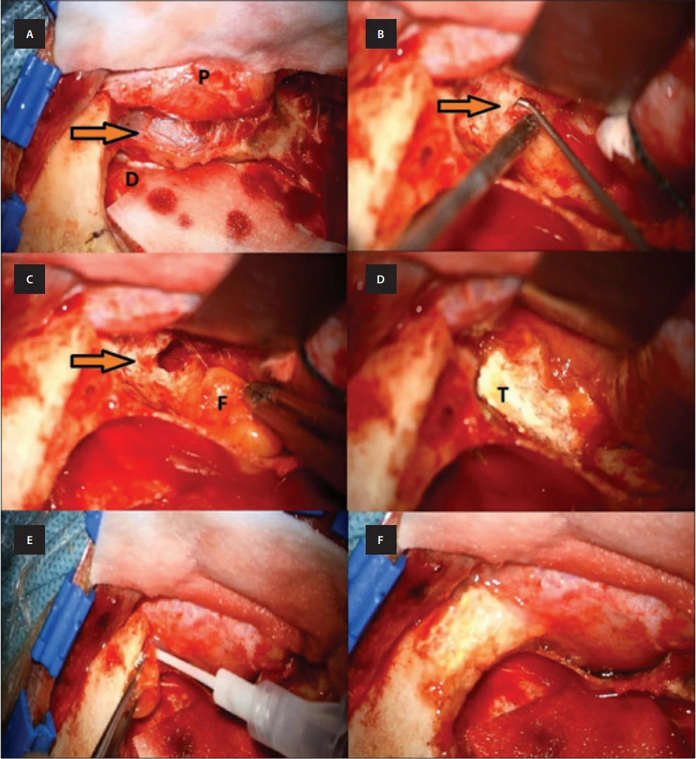
A 49-year-old female was referred to our department with downward deviation of the globe and exophthalmos on the right, without pain or oculomotor dysfunction. Until then, the patient had been monitored for a progressive intraorbital process for more than 10 years at a different institution, without any imaging studies. The patient had sustained a head injury 17 years earlier (she was hit by a motorcycle as a pedestrian). A CT scan was not performed at that time. However, as a consequence of the injury, the patient had prolonged swelling of the right eye, without CSF leakage. A subsequent CT scan of the orbit and paranasal sinuses revealed a well-encapsulated cystic lesion with a calcified wall in the right extraconal region, measuring 2,3 × 2,8 × 1,5 cm, compressing the globe and the superior rectus muscle (Fig. 1). A defect in the ethmoid sinus (Fig. 1) was visualized on CT bone scans. MRI of the orbit revealed an expansive lesion. On fluid attenuated inversion recovery (FLAIR) sequences, the lesion showed hyperintense signal and compressed superior rectus muscle beneath the orbital roof (Fig. 1) and showed no convincing enhancement on post-contrast MRI (Fig. 1). Based on clinical symptoms and imaging findings, the diagnosis of orbital mucocele was established, and surgical intervention was indicated. We performed a modified one-piece orbitozygomatic (OZ) craniotomy. During separation of the periorbita, a pus-like liquid was released. After removal of the bone flap, a partially calcified cavity lined with epithelium was observed (Fig. 2). The epithelium was removed and sent for histological examination. The walls of the partially calcified cavity were drilled out, while the periorbita was preserved. In accordance with preoperative CT findings, during revision of the cavity we identified a defect in the ethmoid sinus, from which the suspected mucocele originated (Fig. 2). Finally, revision of the ethmoid and frontal sinus defect following OZ craniotomy was performed (Fig. 2). The paranasal sinus defect was obliterated with fat tissue, glue, and TachoSil (Takeda, Tokyo, Japan). The dura mater remained intact. Histologically, the cyst walls were lined with pseudostratified ciliated columnar respiratory epithelium without atypia. The cyst wall was fibrous, with microfragments of bone tissue and no significant inflammatory infiltration. Microbiological culture of the specimen revealed Cutibacterium acnes, which is part of the normal flora of mucosal surfaces [2]. Therefore, the diagnosis of mucocele was confirmed. The postoperative course was uneventful. The patient was discharged on postoperative day 5, and the proptosis regressed after surgery. A postoperative MRI performed six months after surgery confirmed complete removal of the mucocele, with no signs of recurrence.
Skull base fractures, though often asymptomatic, may lead to serious complications such as neurovascular injury, tissue contusions and CSF leakage [3]. Orbital mucoceles represent a rare, slow-growing pathology that may develop as a late complication of trauma, including paranasal sinuses fractures [4]. In general, a mucocele is a tumor-like lesion lined by respiratory mucosa, typically found in the paranasal sinuses and filled with mucus [5]. The most common mechanism of mucocele formation after trauma include obstruction of the sinus drainage pathway and entrapment of mucosal membrane within fracture lines. As these trapped cells continue to produce mucus, a cyst may develop, which can subsequently lead to remodeling of the bony walls or even expansion beyond the sinus boundaries. The development of a mucocele following trauma may not occur immediately, as it can take time for mucus to accumulate and cause noticeable symptoms, as seen in our case. We believe that the latter mechanism was responsible for the formation of the orbital mucocele in our patient, as migration and proliferation of respiratory epithelial cells from the ethmoid sinus into the orbit at the time of skull base fracture may have occurred. However, as the patient did not undergo CT imaging at the time of trauma, the exact origin of the mucocele in our case remains debatable. Nevertheless, the history of trauma together with later imaging findings makes this hypothesis the most plausible explanation.
The symptomatology of orbital mucocele is typically characterized by exophtalmos, globe displacement, and oculomotor dysfunction, accompanied by diplopia or altered vision when significant compression of optic structures is present [6]. MRI signal intensity is variable and depends on the relative proportions of water, mucus and protein [7]. Treatment of mucocele is complete surgical removal, including the epithelial lining, to minimize the risk of recurrence from residual mucosa [8]. In our case, the orbital mucocele was removed via a modified one-piece OZ approach, which was selected due to the size of the lesion and presence of calcified walls requiring drilling. Compared with endoscopic approaches, we believe OZ craniotomy provided better exposure and facilitated management of the defect.
A literature review was conducted using the PubMed database to identify relevant English-language articles by using combinations of search terms and synonyms for “mucocele”, “orbital mucocele”, “sequela of skull base trauma”, and “mucocele after skull base trauma” through August 2025. To the best of our knowledge, our case represents only the second reported instance of an isolated orbital mucocele developing as a late sequela following skull base fracture in the English-language literature. The only previously published case was reported by Rajabi et al. in 2012, where the mucocele arose exclusively within the orbit, without communication with the paranasal sinuses [9].
Isolated orbital mucocele may develop as a rare complication following a skull base fracture. When present, complete surgical removal of mucocele, including the epithelial lining, along with revision of the underlying bone defect, is recommended. Similar to other late complications following skull base fractures, a mucocele may develop and become symptomatic several years after the initial injury, as demonstrated in our case. This highlights the importance of long-term follow-up in selected patients after skull base trauma.
Conflict of interest
The authors declare they have no potential conflicts of interest concerning drugs, products, or services used in the study.
Sources
1. Lund VJ, Rolfe ME, Howard DJ. Sphenoidal sinus mucocele: a report of 30 cases. J Laryngol Otol 1998; 112 (1): 27–30. doi: 10.1017/S0022215109991551.
2. Achermann Y, Goldstein EJ, Coenye T et al. Propionibacterium acnes: From commensal to opportunistic biofilm-associated implant pathogen. Clin Microbiol Rev 2014; 27 (3): 419–440. doi: 10.1128/CMR.00092-13.
3. Baugnon KL, Hudgins PA. Skull base fractures and their complications. Neuroimaging Clin N Am 2014; 24 (3): 439–465. doi: 10.1016/j.nic.2014.03.001.
4. Har-El G. Mucoceles of the paranasal sinuses: diagnosis and management. Otolaryngol Clin North Am 2001; 34 (1): 243–268.
5. Courson AM, Stankiewicz JA, Lal D. Contemporary management of paranasal sinus mucoceles: a meta-analysis. Laryngoscope 2014; 124 (2): 378–386. doi: 10.1002/lary.24309.
6. Herndon M, McMains KC, Kountakis SE. Presentation and management of extensive fronto-orbital-ethmoid mucoceles. Am J Otolaryngol 2007; 28 (3): 145–147. doi: 10.1016/j.amjoto.2006.07.010.
7. Rao VM, el-Noueam KI. Sinonasal imaging: anatomy and pathology. Radiol Clin North Am 1998; 36 (5): 921–939. doi: 10.1002/alr.21178.
8. Scangas GA, Gudis DA, Kennedy DW. The natural history and clinical characteristics of paranasal sinus mucoceles: a clinical review. Int Forum Allergy Rhinol 2013; 3 (9): 712–717.
9. Rajabi MT, Hosseini SS, Tabatabaie SZ et al. Isolated intraorbital mucocele: a rare case report. Iran J Ophthalmol 2012; 24 (1): 57–60.
Labels
Paediatric neurology Neurosurgery NeurologyArticle was published in
Czech and Slovak Neurology and Neurosurgery

2026 Issue 5
Most read in this issue
- Safety aspects of transitioning from alteplase to tenecteplase in the treatment of acute ischemic stroke – a narrative review
- 100 years of the Czech Department of Neurology, Charles UniversityThe fates of Czech and German neurologists in Prague from 1938 to 1945
- Anatomy, etiology, and graphic imaging findings of hypertrophic olivary degeneration – description of 10 cases
- Patterns of cognitive-motor interference in patients with post-stroke cognitive impairment